Skip to content

Air Embolism in the Cath Lab: Coronary, Pulmonary and Cerebral Complications
Practical guide for cardiologists to recognise, manage, and prevent air embolism in the cath lab – focusing on coronary, pulmonary and cerebral circulation.
This article is written for interventional cardiologists, fellows and cath lab staff who want a concise, evidence-informed overview of air embolism in the cath lab.
For more cath lab topics, visit our Cath Lab Complications section or learn more about Dr A M Thirugnanam.
What Is Air Embolism?
An air embolism occurs when gas enters the vascular system and obstructs blood flow. Even small volumes can cause critical ischemia when they reach the proximal LAD, main pulmonary artery, or cerebral arteries.
- Acute myocardial or cerebral ischemia
- Ventricular arrhythmias and conduction block
- Hemodynamic collapse or cardiac arrest
- Neurologic injury including stroke and seizures
In the cath lab, air embolism is almost always iatrogenic and related to:
- Incomplete de-airing of catheters, sheaths and manifolds
- Loose connections or open stopcocks
- Empty contrast or saline bags (“air run”)
- Transseptal and structural interventions with large-bore sheaths
For additional background on gas embolism, see the overview on venous and arterial gas embolism (UpToDate – subscription) or the open-access review on air embolism in interventional procedures (PubMed Central) .
1. Coronary Air Embolism
Typical Clinical Scenario
Coronary air embolism usually presents during diagnostic angiography or PCI as:
- Sudden, severe chest pain immediately after contrast injection
- New ST-segment elevation or depression on ECG
- Hypotension, bradycardia, high-grade AV block, VT or VF
- Angiographic “bead-like” lucent columns in the coronary lumen with abrupt loss of flow

Stepwise Coronary Management in the Cath Lab
The goal is rapid removal or dispersion of air and restoration of TIMI 3 flow.
1. Stop Further Injections
- Clearly announce: “Air embolism – stop all injections.”
- Stop contrast and saline injections through the affected line.
- Close all stopcocks and inspect the manifold and syringes for air.
2. Administer 100% Oxygen
- Provide 100% oxygen (FiO2 1.0) via mask or ventilator.
- High FiO2 accelerates nitrogen washout and bubble shrinkage.
3. Guide Catheter Aspiration
- Deeply seat the guiding catheter in the affected coronary ostium.
- Attach a 20–30 ml syringe and aspirate forcefully; discard each aspirate.
- Repeat multiple times until air is no longer visualised angiographically.
4. Distal Aspiration
- If distal bubbles remain or flow is TIMI 0–1, advance an over-the-wire balloon, microcatheter, or aspiration catheter to the affected segment.
- Aspirate small volumes repeatedly to clear distal air.
5. Vasodilators for Spasm and No-Reflow
- Give intra-coronary nitroglycerin, verapamil/diltiazem, and/or adenosine.
- These help relieve secondary spasm and microvascular obstruction.
6. Hemodynamic and Rhythm Support
- Support blood pressure with IV fluids and vasopressors/inotropes as needed.
- Use temporary pacing for severe bradycardia or high-grade AV block (especially RCA air).
- In massive embolism with cardiogenic shock, consider IABP, Impella or ECMO in experienced centres.
For related content, see our article on no-reflow phenomenon after PCI .
2. Pulmonary (Venous) Air Embolism
Pulmonary air embolism arises from the venous side of the circulation, often due to:
- Central venous line placement or removal
- Large-bore venous sheaths used for structural heart procedures
- Loose or disconnected high-flow infusion lines
Clinical Presentation
- Sudden dyspnea, cough or pleuritic chest pain
- Hypoxia and hypotension
- Tachycardia; rarely, a “mill wheel” murmur

Management in the Cath Lab
1. Stop the Source of Air
- Immediately clamp or disconnect the offending venous line or sheath.
- Confirm that no further air can be entrained into the venous system.
2. Durant Maneuver (Positioning)
- Place the patient in left lateral decubitus and Trendelenburg position.
- This helps trap air in the RV apex and reduce further embolisation to the pulmonary artery and, in the presence of a shunt, systemic circulation.
3. Aspiration from the Right Heart (If Feasible)
- If a right atrial or right ventricular catheter (e.g. Swan-Ganz) is present, position the tip in RA/RV where air pools.
- Aspirate repeatedly with a large syringe to remove as much air as possible.
4. Support Right Ventricular Function and Oxygenation
- Administer 100% oxygen.
- Maintain adequate preload with IV fluids.
- Use vasopressors for hypotension and to support systemic perfusion.
- Consider inhaled nitric oxide for RV afterload reduction in selected cases.
- In refractory shock or arrest, VA-ECMO may be lifesaving in experienced centres.
More information on venous air embolism is available in the review from major cardiology journals (AHA / Circulation) .
3. Cerebral (Systemic) Air Embolism
Cerebral air embolism is uncommon but potentially devastating. In the cath lab, risk is increased during:
- Transcatheter aortic valve implantation (TAVI)
- Left atrial appendage closure and other LA/LV structural procedures
- MitraClip and transcatheter mitral interventions
- Paradoxical embolism via patent foramen ovale (PFO) or atrial septal defect (ASD)
- Aortic manipulation with air tracking into arch vessels
Clinical Presentation
- Sudden loss of consciousness
- New focal neurological deficit (e.g. hemiparesis, aphasia, visual loss)
- Seizures or status epilepticus
- Failure to wake as expected after the procedure


Management Priorities
1. Stabilise and Oxygenate
- Secure the airway where indicated and provide 100% oxygen (FiO2 1.0).
- High FiO2 shrinks bubbles and improves oxygenation of ischemic brain tissue.
2. Maintain Cerebral Perfusion
- Avoid hypotension; support blood pressure with IV fluids and vasopressors.
- Maintain adequate cerebral perfusion pressure to reduce secondary injury.
3. Urgent Neurology and Hyperbaric Referral
- Involve the stroke/neurology team early.
- Hyperbaric oxygen therapy (HBOT) is the key advanced treatment for significant cerebral air embolism.
- Arrange rapid transfer to a hyperbaric centre where appropriate and available.
4. Imaging
- Obtain CT or MRI of the brain to confirm the diagnosis and exclude hemorrhage.
- Recognise that air can re-dissolve quickly; a negative early scan does not fully exclude air embolism.
For an overview of hyperbaric indications, see the information from the Undersea and Hyperbaric Medical Society (UHMS) .
4. Imaging Layout for Teaching and SEO
For teaching sessions, conference slides, and SEO-optimised medical blogs, structure your images clearly:
- Coronary air embolism: angiographic frame with labeled lucent bubbles and TIMI flow grade (Figure 1).
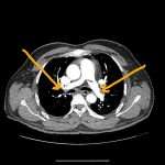
- Pulmonary air embolism: CT chest with arrows marking intraluminal air (Figure 2).
- Cerebral air embolism: brain CT/MRI with labels over cortical air foci (Figures 3 and 4).
When editing the images in Canva or PowerPoint, add simple overlays such as “Coronary”, “Pulmonary” and “Cerebral” to improve clarity and on-page engagement.
5. Prevention: A Zero-Tolerance Strategy for Air in the Cath Lab
Prevention is the most powerful strategy against air embolism in the cath lab. A zero-tolerance approach to visible and microbubbles includes:
- Meticulous de-airing of catheters, syringes, manifolds and tubing
- Never allowing contrast or saline bags to run dry
- Back-bleeding and flushing every new catheter before engaging the vessel
- Submerging structural devices in saline while loading and gently tapping out microbubbles
- Using a standardised checklist and regular team drills for air embolism recognition and response
You can integrate these steps into your local cath lab air embolism checklist and fellowship training curriculum.
6. Conclusion: Key Takeaways for Interventional Cardiologists
In the cath lab, seconds matter. Recognising the classic patterns of air embolism — chest pain and ST changes immediately after injection, sudden hypoxia after venous manipulation, or unexpected neurological deficit during structural intervention — should trigger a rehearsed response:
- Stop the source of air
- Aspirate through the appropriate catheter (coronary, RA/RV, or device sheath)
- Administer 100% oxygen
- Stabilise hemodynamics and treat arrhythmias
- Escalate early to HBOT, mechanical circulatory support or ECMO when indicated
With disciplined technique, a vigilant team and clear cath lab protocols, air embolism can be converted from a catastrophic surprise into a rare, manageable, and teachable event.
To explore more topics in interventional cardiology, visit our Interventional Cardiology Blog and subscribe for updates on new procedures, case discussions, and conference highlights.