
Stent thrombosis is the one complication that turns a technically satisfying case into a catastrophe within hours. Contemporary drug-eluting stents have pushed the overall rate to roughly 0.5% of PCI cases, yet 30-day mortality after an event still approaches 25%. The rate fell; the lethality did not.

- Why stent thrombosis still kills
- Decision 1: The first ten minutes
- Decision 2: What the angiogram hides
- Decision 3: The imaging run that changes management
- Decision 4: Bailout pharmacology at the table
- Decision 5: Getting the stent right the second time
- Decision 6: Discharge therapy and preventing recurrence
- Frequently asked questions
Why stent thrombosis still kills
Every registry tells the same story. Mortality after stent thrombosis climbs from about 14.6% at one year to 33.8% at ten years. Within 60 days of the index event, cardiac death runs at 19.5% and reinfarction at 17.9%. Recurrent stent thrombosis is not rare either — roughly one in eight patients has a second event.
The reason is mechanical. A thrombosed stent occludes a vessel that has already been prepared, dilated and denuded of its endothelium. There is no collateral recruitment, no ischaemic preconditioning, no warning angina. The territory dies at the speed of the occlusion.
That is why I treat every suspected stent thrombosis as a primary PCI activation, not a diagnostic dilemma. The pre-test probability in a patient with chest pain and ST elevation days after PCI is high enough that waiting for troponin is indefensible.
Decision 1: The first ten minutes
Three things happen simultaneously: a 12-lead ECG, a loading dose, and a phone call to the lab. The ECG usually shows ST elevation in the stented territory, but a subacute event in a diagonal or an OM can present with nothing more than reciprocal change and a hypotensive, clammy patient.
Loading matters more than most units acknowledge. Aspirin 150–325 mg if not already on board, plus a potent P2Y12 inhibitor — ticagrelor 180 mg or prasugrel 60 mg. I avoid re-loading clopidogrel in this setting. If the patient thrombosed a stent while taking clopidogrel, clopidogrel has already answered the question about its adequacy.
Anticoagulation is unfractionated heparin to an ACT of 250–300 seconds, or bivalirudin where bleeding risk dominates. In a cardiogenic-shock presentation I prefer heparin for its predictability and reversibility.
The history that predicts the mechanism
Two questions decide much of what follows. When did the patient last take the P2Y12 inhibitor, and why did they stop? Median time to thrombosis after clopidogrel cessation within the first six months is about nine days. A patient who stopped for a dental extraction eight days ago is a pharmacological problem. A patient with impeccable adherence is a mechanical one, and the imaging catheter becomes mandatory.
Decision 2: What the angiogram hides
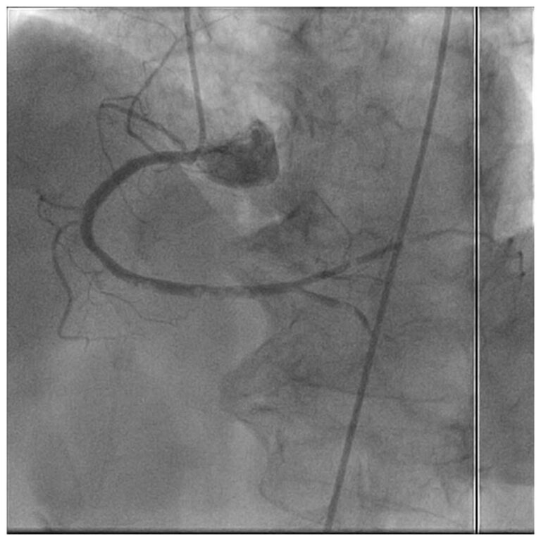
The angiogram confirms the occlusion and nothing else. Luminal silhouette cannot show you stent underexpansion, which carries roughly a thirteen-fold relative risk increase for early stent thrombosis. It cannot show malapposition, present in 18.1% of stent thrombosis cases. It cannot reliably grade an edge dissection.
Underexpansion accounts for about 26% of early events. Overlapping stents feature in 36.0% of cases versus 24.7% of controls, bifurcation lesions in 45.5% versus 36.5%. These are all findings the angiogram either misses or underestimates once thrombus fills the frame.
My working rule: if the angiogram alone were sufficient, the first procedure would not have failed.
Decision 3: The imaging run that changes management
I restore flow first — wire, gentle balloon, aspiration if thrombus burden is large — then image. Imaging into a fully occluded segment wastes contrast and time.
Once TIMI 2–3 flow returns, OCT or IVUS answers the only question that matters: is this a mechanical failure I can fix now, or a pharmacological failure I must fix on the ward? RENOVATE-COMPLEX-PCI showed imaging-guided PCI reduces the composite of cardiac death, target-vessel MI and revascularisation against angiographic guidance alone. ADAPT-DES linked IVUS guidance to lower one-year definite or probable stent thrombosis across 9,961 patients.
The targets I hold myself to are a minimum stent area of at least 5.0 mm² on IVUS or 4.5 mm² on OCT, full strut apposition across the treated segment, and no untreated edge dissection. Calcium features that predict underexpansion — arc ≥180°, thickness ≥0.5 mm, length >5 mm — should have been addressed at the index procedure and, if missed, must be addressed now.
Decision 4: Bailout pharmacology for stent thrombosis at the table
Oral loading in a vomiting, hypoperfused patient has unreliable absorption. This is where intravenous agents earn their place.
Cangrelor gives immediate, titratable P2Y12 blockade with offset within an hour. A glycoprotein IIb/IIIa inhibitor — tirofiban or eptifibatide — remains a reasonable choice when thrombus burden is heavy and bleeding risk is acceptable. For microvascular obstruction after flow restoration, intracoronary adenosine 60–200 mcg through the guide, or nicorandil where available, is my default.
Aspiration thrombectomy deserves a measured position. It reduces distal embolisation but has not reduced MACE in randomised data, and routine use in the TOTAL trial carried a stroke signal. I use it selectively for large, visible thrombus burden — not reflexively because the lesion looks messy.
Decision 5: Getting the stent right the second time
Restoring flow is not the endpoint. If underexpansion caused the event, high-pressure post-dilatation with a non-compliant balloon sized to the media-to-media diameter is the treatment. If an edge dissection propagated, it needs covering. If strut malapposition is focal and the lumen is adequate, aggressive re-stenting adds metal without adding benefit.
I resist the instinct to implant another stent for its own sake. More layers of metal in a segment that has already sustained stent thrombosis increases strut burden, delays healing and sets up the next event. Strut thickness alone matters — struts above 162 µm are around 1.5 times more thrombogenic than thin struts near 81 µm.
Re-stent when there is dissection you cannot leave, a gap in coverage, or a lumen that will not expand. Otherwise optimise what is already there.
Decision 6: Discharge therapy and preventing recurrence
Aspirin continues indefinitely. Dual antiplatelet therapy runs at least 12 months and I extend it in most of these patients, because a prior stent thrombosis is itself among the strongest predictors of the next one. The potent P2Y12 inhibitor stays; de-escalation to clopidogrel in someone who has already thrombosed on it makes no sense.
Where the stent thrombosis occurred despite documented adherence to a potent agent, I look further: platelet function testing, CYP2C19 genotype if clopidogrel is unavoidable, a proton-pump interaction review, thrombocytosis above 400 K/ml, and an underlying malignancy or hypercoagulable state. A systemic immune-inflammation index at or above 636 independently predicts events and costs nothing to calculate.
Then the unglamorous work. High-intensity statin, guideline-directed medical therapy, glycaemic and renal optimisation, smoking cessation, and a conversation about DAPT interruption that the patient will actually remember. Insulin-treated diabetes carries early event rates of 1.7% against 0.9%; an eGFR below 30 predicts the late events.
What I tell the patient before discharge
One sentence, repeated until it lands: do not stop either tablet for any reason without calling this number first — not for a dental appointment, not for endoscopy, not for a skin lesion. Then I write it on the discharge summary in bold and send a copy to the referring physician.
Frequently asked questions
How quickly does stent thrombosis occur after PCI?
Acute events happen within 24 hours and subacute events between 24 hours and 30 days. Late events occur from 31 days to one year, and very late events beyond one year. Acute and subacute events carry the highest early mortality because the myocardium at risk has no collateral protection.
Should intravascular imaging be used in every case?
In my practice, yes, once flow is restored. Imaging distinguishes a correctable mechanical cause from a pharmacological failure, and that distinction changes both the procedure and the discharge prescription.
Is aspiration thrombectomy recommended?
Selectively. It reduces distal embolisation with large thrombus burden but has not improved hard outcomes in randomised trials, and routine use carries a stroke signal. Reserve it for heavy, angiographically evident thrombus.
Which P2Y12 inhibitor after an event on clopidogrel?
Ticagrelor or prasugrel, unless there is a specific contraindication. An event that occurred on clopidogrel is prima facie evidence of inadequate platelet inhibition in that patient.
How long should dual antiplatelet therapy continue?
At least 12 months, and longer in most survivors of an event. Extended therapy reduces ischaemic events at the cost of bleeding, so the decision is individualised using bleeding risk and the DAPT score.
Read next: Cardiology Learning Center · Cardiology Mentorship Programme · Clinical article index
References. Stent Thrombosis: A Contemporary Guide to Definitions, Risk Factors, and Management. Front Cardiovasc Med 2025;12:1622235. · 2025 ACC/AHA/ACEP/NAEMSP/SCAI Guideline for the Management of Patients With Acute Coronary Syndromes. Circulation 2025. · RENOVATE-COMPLEX-PCI. · ADAPT-DES. · TOTAL trial angiographic core laboratory analysis, EuroIntervention.
This clinical review of stent thrombosis is intended for medical professionals and does not replace individual clinical judgement.
Discover more from Dr AM Thirugnanam
Subscribe to get the latest posts sent to your email.