
An ostial left main wrapped in 360-degree calcium is the lesion that makes me slow down before I scrub. Below are the seven decisions I will make for a woman whose case I am planning now, in the order I make them on the table. This is operator reasoning anchored to the evidence, not a procedural report.
The case: a calcified ostial left main in a 75-year-old woman
She is 75, with an ejection fraction of 60%. Her mid-RCA has already been treated with PCI, and that vessel becomes her lifeline whenever I occlude the left main. Angiography and IVUS show circumferential calcium from the aorto-ostium to the distal left main, reference diameter about 4.5 mm, with an 80% mid-LAD lesion and a 90% proximal LCx lesion.
In EXCEL and NOBLE, PCI performed comparably to CABG for isolated ostial or shaft disease with low SYNTAX. Hers is left main plus two vessels after RCA PCI, so the heart team weighs her individually; I assume PCI. No randomised IVL-versus-rotablation trial has enrolled the left main. Our ostial left main data are registries, case reports and a 148-patient systematic review of mostly distal disease.
Decision 1: Image the ostial left main with IVUS, not OCT
OCT needs a sealed column of flush; with the guide tip at or outside a true aorto-ostial lesion, blood washes back and the image cannot be trusted. IVUS has no such dependence, so it is my first run.
From the pullback I want calcium arc, length, minimum lumen area and the position of the tightest calcified rings, where fracture matters. IVUS cannot see behind calcium, so I judge severity by arc and length.
Decision 2: Choose the calcium-modification device
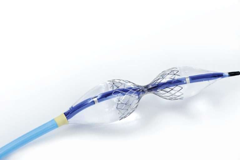
For a crossable lesion with concentric calcium, IVL comes first: lowest perforation and slow-flow risk, no burr to entrap, and fractures that reach deep calcium. In an elderly woman with a large calcified ostial left main, that settles it.
ICARE OFDI randomised 169 patients with crossable calcium to rotablation or IVL. Minimum stent area was 6.0 ± 2.3 mm² with IVL and 5.9 ± 2.2 mm² with rotablation, non-inferior within a 0.75 mm² margin. Major strut malapposition was commoner after rotablation (80.2% versus 57.8%, p=0.002), and 12-month target lesion failure was 1.2% with rotablation versus 2.4% with IVL (p=0.61).
The left main was excluded, and rotablation remains indispensable for uncrossable lesions. ROLLER COASTR-EPIC22 agreed in vessels of 2.5–4.0 mm: OCT stent expansion was 86.4% ± 14.1% with rotablation and 85.6% ± 13.3% with IVL (P=0.77), while excimer laser, at 80.3%, failed non-inferiority.
| What I find | My device | Why |
|---|---|---|
| Crossable, concentric 360-degree calcium | IVL first | Safest profile; deep fractures |
| 1.5 mm balloon will not cross or dilate | Rotablation, 1.25–1.5 mm burr, then IVL | Opens a channel for IVL |
| Eccentric calcified nodule | Rotablation, then NC or OPN | IVL is weaker on nodules |
| Aorto-ostial left main | No orbital atherectomy | Crown cannot orbit safely; eccentric sanding |
| Uncrossable lesion or underexpanded stent | Excimer laser, bail-out only | Failed non-inferiority |
Two more trials matter. In PREPARE-CALC, strategy success was 98% with rotablation against 81% with modified balloons (relative risk of failure 9.5, 95% CI 2.3-39.7). ECLIPSE found orbital atherectomy no better than balloon preparation, with minimum stent area 7.67 versus 7.42 mm² (p=0.08).
Decision 3: Rehearse with an undersized NC test inflation
Before IVL, I rehearse it with a 3.0 × 8–12 mm NC balloon (2.5 mm if tight), taken to nominal and no higher than about 12 atm. It stays up 15–20 seconds, one IVL cycle, with the guide backed into the aorta, while the team calls the MAP nadir, ST change, rhythm, symptoms and recovery time.
Stable pressure and recovery inside 30–60 seconds mean IVL without support; a waist at 12 atm confirms lithotripsy is needed. Not a cutting balloon: slow inflation lengthens occlusion, blades blur the read-out, and a blade in an ostial left main ring can dissect into the aortic root. It has a later role at a fibrocalcific branch ostium.
Decision 4: Will she tolerate ostial left main occlusion during IVL?
Each IVL cycle occludes the left main for 15–20 seconds, no longer than an NC inflation for stenting, POT or kissing. A patient who can get through left main stenting can get through IVL, provided cycles are short and recovery is complete. EF 60% and a patent RCA favour her; hypotension, LVEDP above about 20 mmHg, significant valve disease, recent MI or damping would not.
How I deliver the cycles
- Recheck the RCA: no residual stenosis, dissection or slow flow.
- MAP above 80–85 mmHg before each cycle; pressors and atropine drawn up; pads on.
- Guide disengaged during pulses, for true aortic pressure and correct seating.
- Wait 30–90 seconds for ST, pressure and symptoms to settle, not just the IFU minimum pause.
- If borderline, about five pulses per inflation, as Salazar, Escaned and colleagues did in two women aged 91 and 94.
- LCx buddy wire; ACT above 250–300 seconds.
The ostial left main literature is small but reassuring. Kosowski and colleagues treated an 85-year-old woman with a calcified unprotected ostial left main using a 3.5 mm C2 catheter, 80 pulses in eight runs, with no haemodynamic instability and no mechanical support.
In the BENELUX-IVL left main analysis, the 59 left main patients were older (median 79 versus 73 years) with lower EF (41% versus 51%), yet technical success was 93.2% versus 89.8% and one-year cardiac death, MACE and TVR were comparable. Support was used in 6.8% versus 2.1%, mostly planned.
A 6F femoral sheath under ultrasound covers bail-out IABP, Impella or VA-ECMO; prophylactic support is not routine here. If the test inflation goes badly, I either use rotablation first (non-occlusive, but slow flow in diffuse left main calcium means global ischaemia) followed by fewer IVL cycles, or IVL under Impella CP after iliofemoral CT.
Decision 5: Sizing IVL in a 4.5 mm ostial left main
The largest coronary IVL catheter, the Shockwave C2+ 4.0 × 12 mm, gives a ratio near 0.9; the IFU advises the largest size when 1:1 is unavailable. IVUS shows where it will meet the calcified rings. I pulse at the labelled 4 atm (rated burst 10 atm); higher-pressure pulsing for apposition is off-label.
I expect two positions: ostial, with 1–2 mm of balloon in the aorta, and the distal body, staying within 80 pulses per segment and 120 per catheter. A 4.0 mm balloon never sits in a proximal LAD under 3.5–4.0 mm; the branches get their own sizes.
Decision 6: Guide catheter, EBU 3.5, 7F, no side holes
An EBU 3.5 gives the backup to cross calcium to the bifurcation and deliver stents to the mid-LAD and LCx; a JL is too weak for the circumflex. I choose 3.5 over 4 because older women tend to have small aortic roots, where a 4 dives deep, dangerous at a calcified ostial left main. I upsize only for a dilated root, keep a JL 4.0 shelved, and avoid an AL1.
7F allows kissing NC balloons, two wires with IVUS, or a 1.75 mm burr; side holes would hide damping and dilute ostial contrast. I engage to image and disengage to treat, never leaving the tip inside the calcified ostium while treating, and a guide extension waits until the left main is prepared. Access: femoral 7F under ultrasound, or radial 7F slender if the artery is about 2.3 mm or more.
Decision 7: Stent optimisation at the aorto-ostial left main
Recoil is the enemy at the ostial left main. Among 321 BENELUX-IVL patients, Oliveri and colleagues found 100% device success in 48 aorto-ostial lesions versus 98.2% in the rest, but residual stenosis under 30% was reached less often (81.3% versus 90.5%, p=0.06), and in-hospital MACE was 4.2% versus 0.7% (p=0.048), all cardiac deaths.
So I confirm expansion with a 4.5 mm NC at high pressure under IVUS, then implant a 4.0–4.5 mm platform DES with 1–2 mm aortic protrusion, post-dilate hard, POT with a 5.0 NC and flare the ostium.
Common IVUS floors are 8.2 mm² in the left main, 6.3 mm² at the ostial LAD and 5.0 mm² at the ostial LCx; Sandesara and colleagues, in distal left main PCI, met them in 96/85/89% with IVL versus 93/93/100% with rotablation. In a 4.5 mm ostial left main, with a reference area near 16 mm², I aim for 12–13 mm².
If the LCx lesion involves its ostium, this is a Medina 1,1,1 bifurcation: I plan DK-crush or culotte, with IVL to a calcified LCx, preparing the left main first. More worked examples sit in my interesting cases series and the cardiology learning centre.
My pre-procedure checklist for an ostial left main case
- Heart team discussion documented and PCI agreed.
- RCA result reviewed: no residual stenosis, dissection or slow flow.
- Haemodynamic screen: blood pressure, LVEDP, valves, recent MI.
- Femoral 7F under ultrasound plus a 6F bail-out sheath.
- EBU 3.5, 7F, no side holes; JL 4.0 on the shelf.
- Baseline IVUS; OCT not relied upon at the ostial left main.
- NC 3.0 test inflation, 15–20 seconds, guide out.
- 4.0 × 12 mm IVL catheter open; 1.25–1.5 mm burrs available.
- Pressors, atropine, pads, ACT 250–300 seconds.
- 4.5 and 5.0 NC balloons; MSA goal 12–13 mm².
References
- Jurado-Román et al. ROLLER COASTR-EPIC22. JACC Cardiovasc Interv. 2025;18:606-618. DOI
- Honton et al. ICARE OFDI. EuroIntervention. 2026;22(15):e808-e819. DOI
- Abdel-Wahab et al. PREPARE-CALC. Circ Cardiovasc Interv. 2018;11:e007415. DOI
- ECLIPSE investigators. Orbital atherectomy versus balloon angioplasty. Lancet. 2025. Link
- Oliveri et al. IVL in left main disease, BENELUX-IVL. Int J Cardiol. 2025;423:132996. DOI
- Oliveri et al. IVL in aorto-ostial lesions. Catheter Cardiovasc Interv. 2024. DOI
- Sandesara et al. IVL versus rotablation in calcified distal left main PCI. Catheter Cardiovasc Interv. 2023. DOI
- Kosowski et al. IVL for calcified unprotected ostial left main. Kardiol Pol. 2022;80(4):489-490. DOI
- Salazar et al. IVL in calcified distal left main. JACC Case Rep. 2019;1(1):46-49. DOI
- Systematic review: IVL for calcified left main. JSCAI. 2023. Link
- Shockwave Medical. C2+ coronary IVL catheter, instructions for use. IFU
Discover more from Dr AM Thirugnanam
Subscribe to get the latest posts sent to your email.