Pregnancy and Heart Diseases

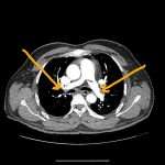
A tremendously increased prevalence of cardiovascular disease (CVD) has been found in women of childbearing age, with the presence of CVD in pregnant women posing a difficult clinical scenario in which the responsibility of the treating physician extends to the unborn fetus. Profound changes occur in the maternal circulation that has the potential to adversely affect maternal and fetal health, especially in the presence of underlying heart conditions. Up to 4% of pregnancies may have cardiovascular complications despite no known prior disease.

In the United States, CVD is the leading cause of death during pregnancy and the postpartum period. It is responsible for 26.5% of all pregnancy-related deaths. Mortality and morbidity associated with CVD are highest among women of color and those with lower incomes.

Physiological Changes during pregnancy

Pregnancy has a profound effect on the circulatory system. Most of these hemodynamic changes start in the first trimester, peak during the second trimester, and plateau during the third trimester. Cardiac output increases by 30-50% secondary to an increase in blood volume and heart rate. Blood pressure decreases by 10-15 mm Hg owing to a decrease in systemic vascular resistance caused by the creation of a low resistance circuit by the placenta and vasodilatation.  Additionally, heart rate normally increases by 10-15 beats per minute. The hematocrit level decreases due to a disproportionate increase in plasma volume that exceeds the rise in red cell mass. 

During the third trimester, cardiac output is further influenced by body position, where the supine position causes caval compression by the gravid uterus. This leads to a decrease in venous return, which can cause supine hypotension during pregnancy. Stroke volume normally increases in the first and second trimester and decreases in the third trimester. This decrease is due to partial vena cava obstruction.

Valvular Heart Disease in Pregnancy

Valvular heart disease in pregnancy is relatively infrequent, with an incidence of less than 1%. In the developed world, valvular disease in women of childbearing age is often congenitally acquired.  Rheumatic heart disease, myxomatous degeneration, previous endocarditis, and bicuspid aortic valves are also encountered. Pregnancy complicated by valvular heart disease tends to have a favorable prognosis if risks are appropriately managed. Management of the pregnant woman with a heart condition requires special expertise, and patients with high-risk conditions should be referred to centers specialized in their care

Safe medications in Pregnancy

 Medications such as hydralazine, methyldopa, digoxin, adenosine, and procainamide can be safely used in pregnancy. 

Contraindicated Medicines in pregnancy

ACE inhibitors, ARB, Amiodarone, and Nitroprusside.

Pregnancy and congenital heart disease

High maternal risk conditions are as follows:

  • The poor functional class before pregnancy (NYHA functional classification II or more) or cyanosis
  • Impaired systemic ventricular function (ejection fraction < 40%)
  • Mitral valve stenosis (area < 2 cm2), aortic valve stenosis (area < 1.5 cm2), left ventricular outflow tract peak pressure gradient greater than 30 mm Hg before pregnancy
  • Preconception history of adverse cardiac events such as symptomatic arrhythmia,  stroke, transient ischemic attack, and pulmonary edema
  • Marfan syndrome
  • Eisenmenger syndrome
  • Pulmonary hypertension

Moderate maternal risk conditions are as follows:

  • Repaired tetralogy of Fallot without significant pulmonic stenosis or regurgitation 
  • Complex congenital heart disease with the anatomic right ventricle serving as the systemic ventricle
  • Mild mitral or aortic valve stenosis
  • Cyanotic lesions without pulmonary hypertension
  • Fontan type circulation
  • Uncorrected coarctation of the aorta

Low maternal risk conditions are as follows:

  • Small ventricular septal defects
  • Atrial septal defects
  • Bicuspid aortic valve without stenosis, regurgitation, or aortic dilation
  • Repaired coarctation of the aorta.
Dr AM Thirugnanam
Dr AM Thirugnanam
Articles: 18

One comment

  1. Your article gave me a lot of inspiration, I hope you can explain your point of view in more detail, because I have some doubts, thank you.

Leave a Reply

Your email address will not be published. Required fields are marked *